Pre-lab Reading
The circulatory system consists of three general parts: a pump (heart), tubes (blood vessels), and fluid (blood). The function of the circulatory system is to exchange gases (especially O2 CO2), transport nutrients, and remove waste products to and from the cells throughout the body.
and
The blood vessels contain and transport all of these materials to and from cells. There are three categories of blood vessels. Arteries carry blood away from the heart to cells and tissues. Veins return blood from the cells to the heart. Capillaries are within the tissues in your body and carry blood between the arteries and veins. The function of the heart is to pump oxygen-rich blood to the cells and oxygen-poor blood to the lungs.
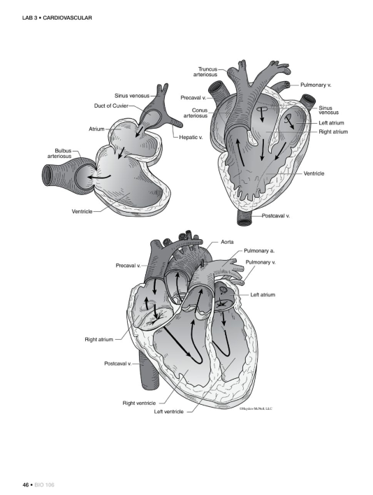
All vertebrate animals have a closed circulatory system, which means that the blood is confined inside of blood vessels. The cardiovascular system of vertebrate animals provides an excellent example of the evolutionary adaptations of different groups of animals in response to natural selection. Aquatic animals and terrestrial animals live in very different environments and thus face different challenges in terms of obtaining oxygen from their environment. Fish have a heart with two chambers and a single circuit for blood flow. Terrestrial animals would have a hard time with a single-circuit system because it wouldn’t provide enough pressure for the blood to move through all of the vessels, whereas the swimming motion and water running across the gills of fish helps to move blood through their blood vessels. Consequently, terrestrial animals evolved a system with double circulation that pumps blood twice, once after the systematic capillaries and again after visiting the lung capillaries, therefore providing more pressure to enable the blood to reach all of the capillaries and return to the heart.
Another difference between different vertebrate taxa is that amphibians and reptiles have hearts with three chambers while mammals and birds have hearts with four chambers. The main reason for the different number of heart chambers relates to the amount of energy required for daily functions for each of the groups. Amphibians and reptiles are ectothermic (they do not produce their own body heat) while mammals and birds are endothermic (they generate their own heat to maintain a steady body temperature). Endothermic animals require much more food and oxygen to sustain the high levels of energy required to keep a constant body temperature. As a result, selection favored a four-chambered heart to provide extra power to pump the blood filled with oxygen and nutrients to cells throughout the body and back again.
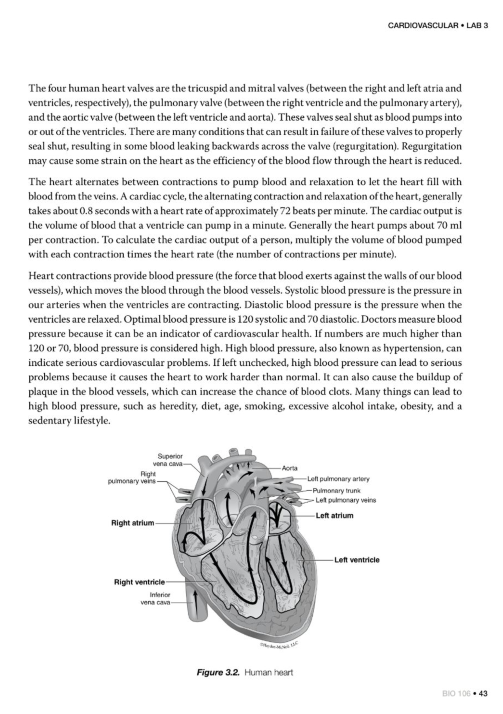
Since the heart is the pump for the whole circulatory system, it is constantly providing the force that moves blood through the blood vessels. The human heart, like those of all mammals, has four chambers, including two atria and two ventricles. The right side of the heart pumps oxygen-poor blood from the body to the lungs, where the carbon dioxide leaves the blood vessels into the lungs and oxygen enters the blood vessels from the lungs. The left side of the heart pumps the oxygen-rich blood to cells throughout the body. The human heart is about the size of your fist and is composed of mostly cardiac muscle tissue. The left and right atria are relatively thin-walled and receive blood returning to the heart from the lungs and the rest of the body, respectively. The two ventricles have thicker walls and pump blood to the lungs and the rest of the body. The left ventricle is thicker and pumps blood throughout the body, while the right ventricle pumps blood to the lungs. The valves between the pulmonary artery (the artery from the heart to the lungs), and the atria and ventricles regulate the blood flow direction. These valves also provide the sound that you hear when you listen to a person’s heartbeat.
The four human heart valves are the tricuspid and mitral valves (between the right and left atria and ventricles, respectively), the pulmonary valve (between the right ventricle and the pulmonary artery), and the aortic valve (between the left ventricle and aorta). These valves seal shut as blood pumps into or out of the ventricles. There are many conditions that can result in failure of these valves to properly seal shut, resulting in some blood leaking backwards across the valve (regurgitation). Regurgitation may cause some strain on the heart as the efficiency of the blood flow through the heart is reduced.
The heart alternates between contractions to pump blood and relaxation to let the heart fill with blood from the veins. A cardiac cycle, the alternating contraction and relaxation of the heart, generally takes about 0.8 seconds with a heart rate of approximately 72 beats per minute. The cardiac output is the volume of blood that a ventricle can pump in a minute. Generally the heart pumps about 70 ml per contraction. To calculate the cardiac output of a person, multiply the volume of blood pumped with each contraction times the heart rate (the number of contractions per minute).
Heart contractions provide blood pressure (the force that blood exerts against the walls of our blood vessels), which moves the blood through the blood vessels. Systolic blood pressure is the pressure in our arteries when the ventricles are contracting. Diastolic blood pressure is the pressure when the ventricles are relaxed. Optimal blood pressure is 120 systolic and 70 diastolic. Doctors measure blood pressure because it can be an indicator of cardiovascular health. If numbers are much higher than 120 or 70, blood pressure is considered high. High blood pressure, also known as hypertension, can indicate serious cardiovascular problems. If left unchecked, high blood pressure can lead to serious problems because it causes the heart to work harder than normal. It can also cause the buildup of plaque in the blood vessels, which can increase the chance of blood clots. Many things can lead to high blood pressure, such as heredity, diet, age, smoking, excessive alcohol intake, obesity, and a sedentary lifestyle.
Online Lab 3: Materials, Methods, and Procedures
Materials
• Cardiio Rhythm smartphone photoplethysmographic (PPG) application
• Stopwatch or timer (must be separate from the smartphone with the PPG app)
• Metal or plastic straw
• Tape
• Balloon or disposable glove
•Scissors
• Glass or cup
Methods
I. Pulse Measurements Before you begin this part of the lab, fill out your hypothesis and prediction in the datasheet.
1. Download the free Cardiio Rhythm PPG app onto your smartphone. Note: You do not need to enable location services on the app for this lab.
2. Use the PPG app to determine your pulse rate at several times during a normal day as a univer-sity student: first thing after you wake in the morning, during a study session, and before falling asleep. Record these numbers.
3. Use the PPG app to determine your pulse rate after sitting for 5 minutes. Record this number.
4. Exercise for 5 minutes (run in place, up and down stairs, around the outside of your house, etc.). Use a timer. Use the app to determine your pulse rate. Record this number.
5. After 30 seconds, and each 30 seconds after that, record your pulse rate again and record it in the datasheet. Continue to record your pulse rate until it is the same as the resting pulse rate.
6. Calculate the recovery index of your pulse rate on the datasheet. 7. Answer the questions in the datasheet.
II. Constructing a One-way Model Valve The valves in your heart control the direction of blood flow by restricting the flow to one direction. Use a straw, balloons or disposable glove, and tape to construct a valve that allows you to blow air through the straw into the water, but does not allow you to draw the water up through the straw into your mouth. There are many solutions to this challenge! Record your results to send to your lab instructor and answer the associated questions in the datasheet.
. Read and Interpret Data—See Part 2 Datasheet on 61 1. Each person in your group should select a different research paper from the list provided by your TA.
2. Find and read the selected paper. 3. Based on the paper that you read, fill in the related questions in the datasheet section.


